

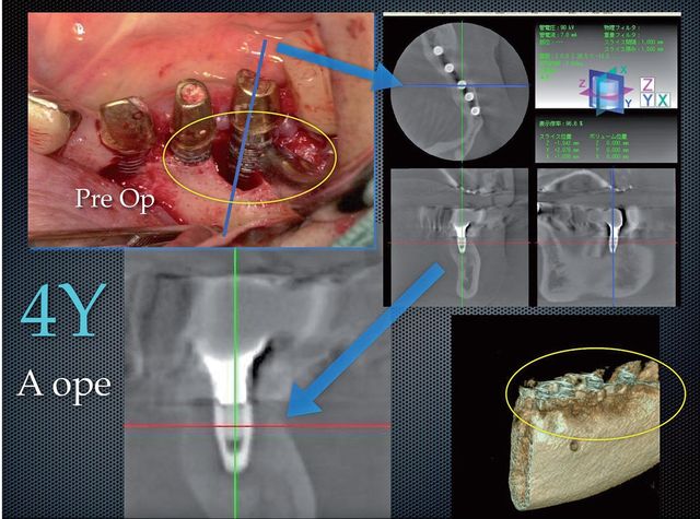
After that, an autogenous bone graft was transplanted and covered with an absorbable membrane, and the site was closed in the conventional way. Figure 3 shows the X-ray taken after 2 years and the X-ray taken after 3 years. All the X-rays show a formation considered to be bone re-growth. Figure 4 shows CT scan image(bucco-lingual cross section) made 4 years after treatment. the area around the implant has stabilized.
Conclusions
The micro-explosions produced by an Er:YAG laser can effectively remove the contaminated titanium oxide layer from an implant failing due to periimplantitis. When accompanied by water spray, irradiation with the Er:YAG laser limits heating of the implant to a few degrees not enough to damage surrounding bone tissue and inhibit osseointegration. These results strongly suggest that the difficulties of treating peri-implantitis can be overcome with Er:YAG laser irradiation.
Correspondence
Atsuhiko Yamamoto
autis@silver.ocn.ne.jp


